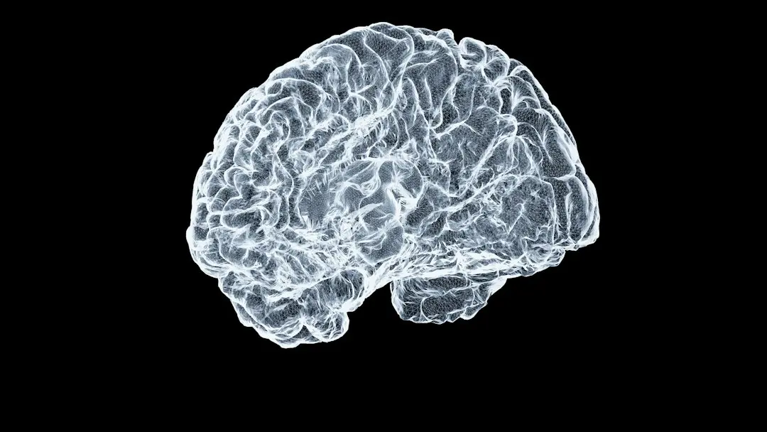
Discovery of a Key Brain Area
Scientists are on the brink of uncovering a new factor contributing to high blood pressure and a method to control it. A researcher from New Zealand and colleagues in Brazil identified a specific brain region that appears to play a significant role in some forms of hypertension. The study, published in 2025, involved specialists from the University of São Paulo and the University of Auckland. The region, known as the parafacial lateral (pFL), can trigger biological changes that increase blood pressure. The pFL is linked to respiratory control, especially forced exhalations during physical effort, coughing, or laughing.
Latest news
 No Military Fix for US‑Iran Tension, Says Tehran’s Foreign Minister
 Stock Futures Hold Steady as Investors Await Fed Minutes and Higher Yields
 Macron's Busy Schedule Threatens His Attendance at COP31 and Undermines Paris Agreement Support
 Dozens detained at Moscow Communist rally as tensions rise over Ukraine drone attacks
Insights from Rodent Experiments
In experiments on rats, researchers observed that the pFL not only regulates breathing but also has an additional function: it can cause blood vessels to constrict. This combination of influencing breathing rhythm and signaling vessels may underpin a form of hypertension. It could explain why about 40 % of patients on antihypertensive drugs still have uncontrolled blood pressure.
Neuronal Mechanisms and Sympathetic Activation
The team hypothesized that neurons in the pFL might link subtle breathing rhythm variations—often imperceptible—to increased sympathetic nervous system activity, the „fight or flight” response that directly affects blood pressure regulation. This idea aligns with earlier research suggesting a connection between blood pressure and brain or nervous system function. The authors emphasized that roughly half of hypertensive patients have a neurogenic component to their disease, making it essential to understand the mechanisms that generate sympathetic excitation.
Genetic Manipulation Confirms the Role
To test their hypothesis, researchers used genetic editing in rats to selectively activate or deactivate pFL neurons. While doing so, they monitored respiratory nerve activity, sympathetic nerve activity, and blood pressure. They found that activating pFL neurons triggered other brain circuits that, in turn, raised blood pressure in the animals. They precisely mapped the communication network between this region and other neurons, comparing the data with healthy rats that did not exhibit hypertension.
Impact on Vascular Constriction
In hypertensive rats, pFL neurons not only supported breathing but actively participated in vascular constriction. This discovery opened a new therapeutic perspective: when blood pressure was high, the pFL became active, and its inactivation lowered blood pressure to normal levels. Physiologist Julian Paton from the University of Auckland highlighted the significance of this intervention.
Another intriguing aspect of the research is the link between the pFL and sleep apnea, a disorder characterized by nighttime breathing problems. Researchers explained that although pFL neurons are not involved in normal breathing, they become active in response to high carbon dioxide or low oxygen levels—conditions typical of sleep apnea episodes. This may explain why people with this disorder have an increased risk of developing hypertension through the same mechanism that connects respiratory control with blood flow regulation.
Connection to Sleep Apnea
The researchers acknowledged that the study was conducted exclusively on animal models. While it is likely that the same neural circuits exist in humans, definitive evidence is still lacking. Nonetheless, the data suggest that about one‑third of the global population suffers from some form of hypertension, and many lack access to effective medication. Developing new therapeutic strategies is therefore an urgent priority, as uncontrolled hypertension significantly raises the risk of serious heart disease and even dementia.
Study Limitations and Future Directions
The team noted that the next step is to design drugs that can selectively target pFL neurons without affecting other neural systems. They have already made progress by studying carotid bodies—clusters of cells in the neck that act as microscopic sensors and can influence pFL neuron activity from outside the brain. These structures may represent a viable therapeutic target for future pharmacological interventions.
Conclusion
This study adds a new dimension to our understanding of hypertension and offers hope for patients who do not respond adequately to existing treatments. By emphasizing the brain’s role in blood pressure regulation and identifying a specific mechanism, researchers are paving the way for innovative therapies that could transform hypertension management worldwide.